The deltoid ligament provides support to the medial part of the ankle (closest to the midline). The calcaneofibular ligament (CFL), which connects the fibula to the calcaneus, or heel bone, also provides lateral support. These ligaments include the anterior talofibular ligament (ATFL) and the posterior talofibular ligament (PTFL). The talus and the fibula are connected by a strong group of ligaments, which provide support for the lateral aspect of the ankle. Together the tibia and fibula form a bracket-shaped socket known as the mortise, into which the dome-shaped talus fits. This articulation (where two bones meet) is primarily responsible for plantarflexion (moving your foot down) and dorsiflexion (moving your foot up). The weight-bearing aspect of the tibia closest to the foot (known as the plafond) connects with the talus. The ankle joint is a highly constrained, complex hinge joint composed of three bones: the tibia, the fibula, and the talus. The ankle region refers to where the leg meets the foot (talocrural region). They occur most commonly in young males and older females. Īnkle fractures are common, occurring in over 1. Significant recovery generally occurs within four months while completely recovery usually takes up to one year. Non-operative treatment includes splinting or casting while operative treatment includes fixing the fracture with metal implants through an open reduction internal fixation ( ORIF). Ankle stability largely dictates non-operative vs. Special X-ray views called stress views help determine whether an ankle fracture is unstable. The Ottawa ankle rule can help determine the need for X-rays.

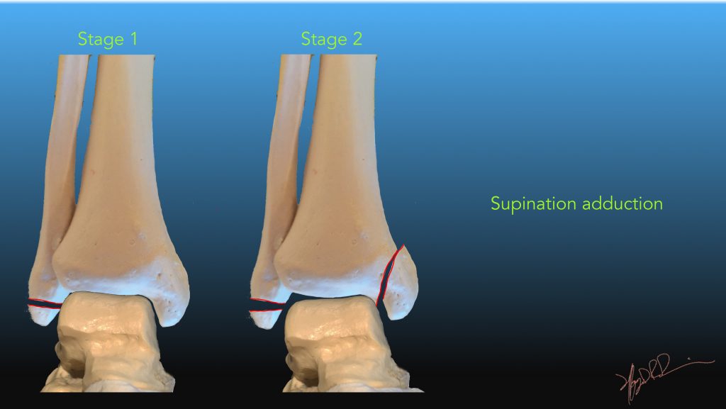
Types of ankle fractures include lateral malleolus, medial malleolus, posterior malleolus, bimalleolar, and trimalleolar fractures.

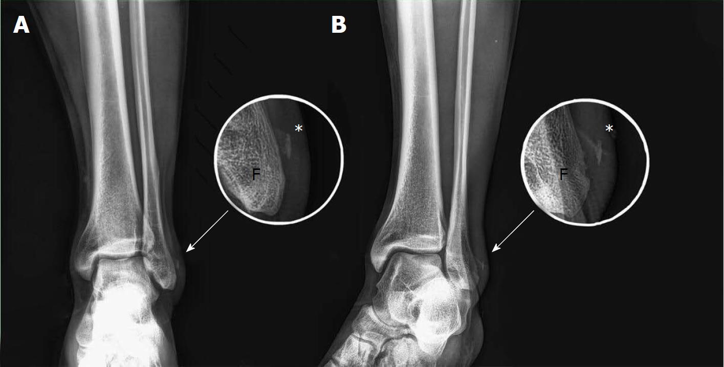
Īnkle fractures may result from excessive stress on the joint such as from rolling an ankle or from blunt trauma. Complications may include an associated high ankle sprain, compartment syndrome, stiffness, malunion, and post-traumatic arthritis. Symptoms may include pain, swelling, bruising, and an inability to walk on the injured leg. Rheumatoid arthritis, gout, septic arthritis, Achilles tendon rupture Īn ankle fracture is a break of one or more of the bones that make up the ankle joint. Lateral malleolus, medial malleolus, posterior malleolus, bimalleolar, trimalleolar High ankle sprain, compartment syndrome, decreased range of motion, malunion Pain, swelling, bruising, inability to walk (1) fibula, (2) tibia, (arrow) medial malleolus, (arrowhead) lateral malleolus Eight weeks after surgery, healing of the pelvic and tarsal fractures, as determined by radiographs, was considered normal and the dog was ambulating with minimal lameness.Fracture of both sides of the ankle with dislocation as seen on anteroposterior X-ray.

The integrity of the tibiotalar short component of the MCL was confirmed during surgical stabilization of the talar fracture. Surgery was performed to repair the talar fracture and sacroiliac luxation. The authors suggest that flexion of the tarsal joint during palpation and radiography stabilized the trochlea of the talus by tensioning the intact tibiotalar short component of the MCL, whereas laxity within the long and tibiocentral MCL components spanning the talus permitted further displacement of the talar neck fracture. 2 However, in the dog reported here, crepitus and instability were caused by the talar fracture. In contrast to the tibiotalar short component, the long and tibiocentral components of the MCL span the talus and are taut in extension but lax in flexion.ĭetection of crepitus and instability only during pronation of a flexed tarsal joint suggests isolated disruption of the tibiotalar short component of the MCL. 2 The tibiotalar short component, a stout ligament passing plantarodistally from the malleolus to the medial trochlea of the talus, is taut in flexion but somewhat lax in extension. The MCL of the tarsal joint is a complex structure consisting of 3 distinct components. 1 Pronation stress on the flexed tarsal joint applies tension to an isolated (tibiotalar short) component of the MCL. Physical examination findings were the authors' rationale for obtaining dorsoplantar flexed pronation and supination stressed radiographic views. The radiographic diagnosis was oblique fracture of the talus with subluxation of the talocalcaneal joint and avulsion fracture of the lateral malleolus. Dorsoplantar flexed pronation and supination stressed radiographic views confirmed the presence of fractures of the talar neck and lateral malleolus and suggested that the medial collateral ligament (MCL) was intact ( Figure 3). Traditional varus and valgus stressed radiographic views with the tarsal joint held in extension were not helpful.


 0 kommentar(er)
